


With advances in biomedical imaging and translational research, Near-Infrared II (NIR-II, 900–1700 nm) imaging has emerged as a promising technology for deep-tissue visualization. It enables deeper tissue penetration, high spatial resolution, and real-time dynamic imaging capabilities in small animal models. However, conventional silicon-based sensors suffer from severe quantum efficiency drop-offs in this spectrum.
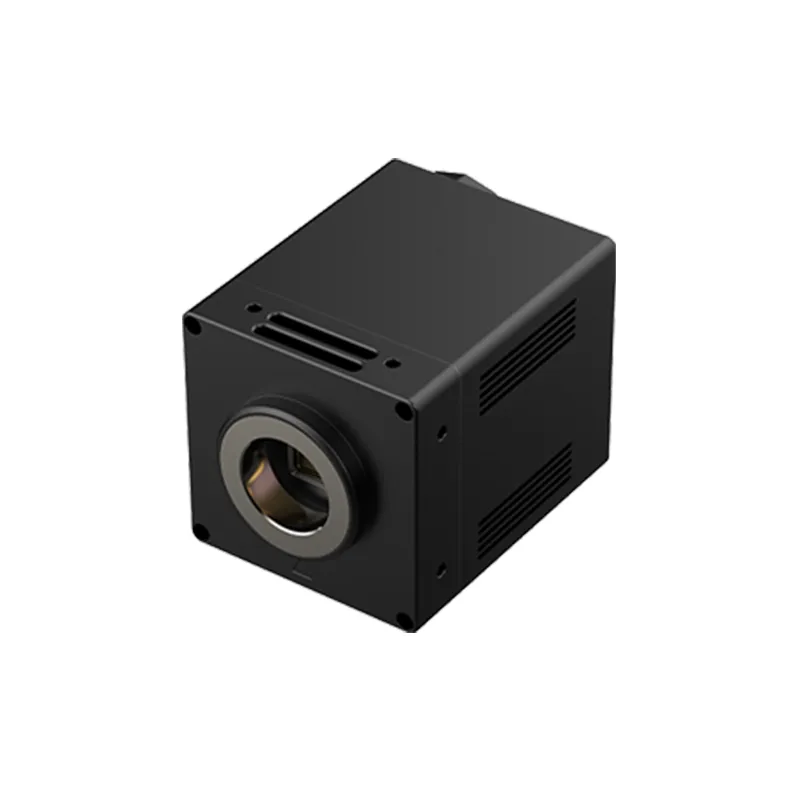
This whitepaper details a robust, validated NIR-II in vivo imaging solution featuring the D-BLUE1 scientific SWIR camera. By leveraging proprietary InGaAs sensor architecture and advanced thermal management, this solution significantly reduces tissue autofluorescence background and achieves high-contrast, deep-tissue visualization of Indocyanine Green in live mice—providing a reliable imaging platform for advanced preclinical and pharmaceutical research. This article focuses exclusively on preclinical research and laboratory imaging applications.
1. What Are the Key Challenges in Conventional Optical Imaging?
1.1 Limited Tissue Penetration in Visible and NIR-I Imaging
The visible light and NIR-I light in the 400–750 nm wavelength range are heavily absorbed and scattered by endogenous biomolecules such as hemoglobin and melanin. This restricts effective imaging depths to a few hundred micrometers, rendering deep internal organs completely invisible to standard optical systems.
1.2 Reduced Imaging Contrast Due to High Background Autofluorescence
Biological tissues emit a strong autofluorescence background under visible and NIR-I excitation. This background noise overlaps with target probe signals, severely degrading the signal-to-noise ratio (SNR) and contrast. Weak signals from deep-tissue targets are easily masked, making stable, long-term tracking difficult.
1.3 Limitations of Silicon-Based Detectors Beyond 900 nm
NIR-II imaging requires detectors with high sensitivity, low dark noise, and superior quantum efficiency (QE) across the 900–1700 nm range. Traditional silicon-based detectors perform well in visible light, but their photoelectric response declines sharply at wavelengths beyond 900 nm, making them incapable of capturing weak NIR-II fluorescence photons.
2. Experimental Validation of D-BLUE1 SWIR Camera for NIR-II In Vivo Imaging Applications
2.1 Experimental Hardware Setup
- Core Detector: Raytron D-BLUE1 scientific-grade InGaAs SWIR camera
- Excitation Source: 793 nm infrared laser
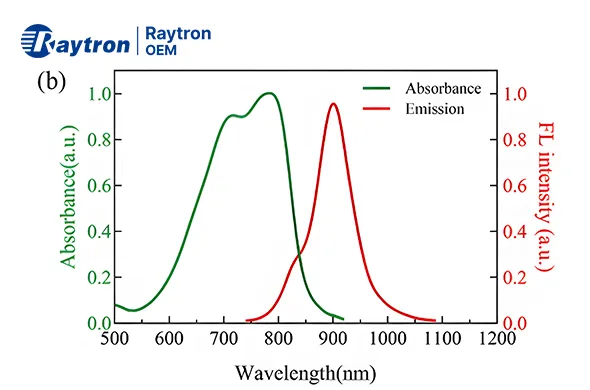
- Fluorescence Probe: Indocyanine green (ICG) (FDA-approved dye optimized for deep-tissue tracking)
2.2 Standard Experimental Workflow
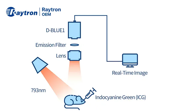
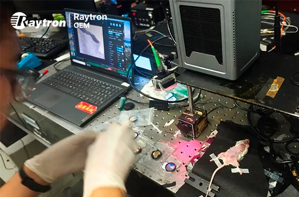
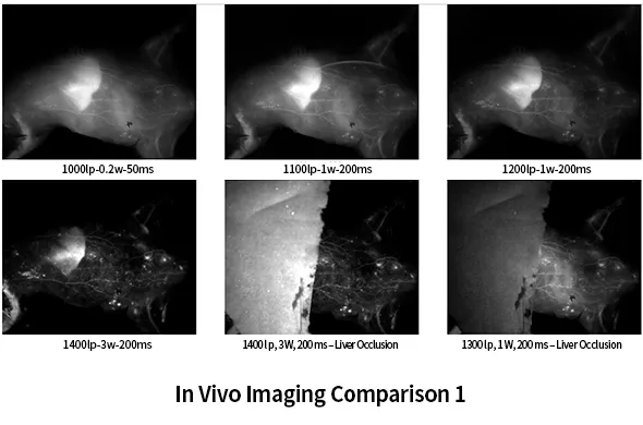
- Subject Preparation: Healthy nude mouse models anesthetized under standard laboratory protocols.
- Administration: Precision retro-orbital vein injection to introduce the ICG tracer into the circulatory system.
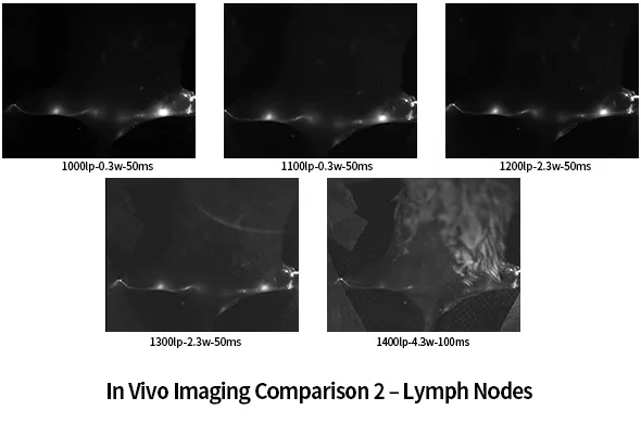
- Data Capture: The D-BLUE1 SWIR camera continuously monitors and captures the dynamic biodistribution and deep-tissue clearance of the probe in real time.
3. What is the D-BLUE1 SWIR Camera
The D-BLUE1 SWIR camera integrates an advanced InGaAs detector with a built-in three-stage thermoelectric cooler (TEC), achieving detector cooling down to -50°C and delivering high-quality imaging performance with low noise.
Designed for scientific and research applications, the camera supports GigE Vision and Camera Link interfaces for flexible connectivity and provides a complete SDK package for secondary development, enabling seamless integration into customized imaging systems.
The D-BLUE1 camera is suitable for a wide range of applications, including:
- Astronomical Observation
Scientific imaging applications require extended spectral response beyond visible wavelengths.
- Spectral Analysis
Raman spectroscopy, microscopic spectroscopy, and other research applications requiring sensitive SWIR detection.
- Semiconductor Failure Analysis
Non-destructive inspection and analysis of semiconductor materials and devices.
- In Vivo Biomedical Imaging
High-sensitivity imaging for biological research and laboratory studies.
4. Advancing Biomedical Research with SWIR Imaging Technology
The D-BLUE1 camera can be used in basic research and preclinical evaluation:
- Oncology & Neurovascular Research: Tumor microenvironment analysis, angiogenesis tracking, and real-time cerebral blood flow/metabolic imaging.
- Biopharmaceutics & Gene Therapy: Stem cell tracking, mRNA/DNA vaccine biodistribution, and high-throughput drug efficacy screening.
- Pathology & Virology: Animal disease model validation, viral mechanisms, and active ingredient screening in traditional medicine/herbal compounds.
Note: The system and technology detailed in this document are strictly designed for fundamental laboratory research and are not intended for human clinical diagnostics.